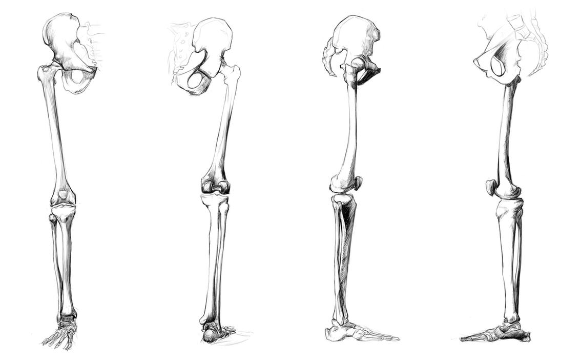
Bones In Leg Diagram - Blank Diagrams - Harvey's A&P / At the distal end of the femur, two rounded condyles meet the tibia and fibula bones of the lower leg to form the knee joint.
Bones In Leg Diagram - Blank Diagrams - Harvey's A&P / At the distal end of the femur, two rounded condyles meet the tibia and fibula bones of the lower leg to form the knee joint.. Together with the upper leg it forms the lower extremity. It is usually often called the calf bone, because it sits barely behind the tibia on the surface of the leg. Bones pain hand and arm bones diagram. Also, defective and old red blood cells are destroyed in bone marrow. The bones of the leg are the femur, tibia, fibula and patella.
Human muscle system the muscles of the. The bones of your leg have roughened patches on their surfaces where muscles are attached. The bones of the leg are the femur, tibia, fibula and patella. The knee is a strong but flexible hinge joint. The bone that goes from your pelvis to your knee is called the femur (say:
Labeled Diagram of the Human Leg by xKeren on DeviantArt from img00.deviantart.net Want to learn more about it? The bones of your leg have roughened patches on their surfaces where muscles are attached. Bones pain hand and arm bones diagram. Master leg and knee anatomy using our topic page. Human anatomy diagrams show internal organs, cells, systems, conditions, symptoms and sickness information and/or tips for healthy living. The knee is a strong but flexible hinge joint. When your muscles contract, they pull the bone they're. Your leg bones are the longest and strongest bones in your body.
This diagram depicts diagram leg bones anatomy.
It mainly serves as an attachment point for the muscles of the lower leg. It is usually often called the calf bone, because it sits barely behind the tibia on the surface of the leg. The foot bones shown in this diagram are the talus, navicular, cuneiform, cuboid, metatarsals and calcaneus. The foot bones shown in this diagram are the talus, navicular, cuneiform, cuboid, metatarsals and calcaneus. Learn vocabulary, terms and more with flashcards, games and other study tools. The knee is a strong but flexible hinge joint. This diagram depicts diagram leg bones anatomy. Together with the upper leg it forms the lower extremity. Leg diagram illustrations & vectors. An intermediate segment, the tibia. The femur, or thigh bone, is the largest, heaviest, and strongest bone in the human body. The human leg consists of 8 bones, 4 per leg. Schema de legs bones diagram diagram showing bones inside human leg ready to jump stock file skeleton of a cat diagram ver 2 svg disposition of rotator cuff muscles diagram.
Want to learn more about it? An electrical wiring diagram can be as simple as a diagram demonstrating how to set up a fresh swap with your hallway. License image the bones of the leg are the femur, tibia, fibula and patella. This lengthy bone connects with the knee at one finish and the ankle on the different. Leg diagram illustrations & vectors.
Anatomy Study - leg bones by Call0ps on DeviantArt from pre00.deviantart.net License image the bones of the leg are the femur, tibia, fibula and patella. An electrical wiring diagram can be as simple as a diagram demonstrating how to set up a fresh swap with your hallway. Normal leg bones are relatively straight, but those affected by paget's disease are porous and curved. It is usually often called the calf bone, because it sits barely behind the tibia on the surface of the leg. The femur, or thigh bone, is the largest, heaviest, and strongest bone in the human body. Your leg bones are very large and strong to help support the weight of your body. The human leg, in the general word sense, is the entire lower limb of the human body, including the foot, thigh and even the hip or gluteal region. Cancellous bone produces red blood cells, platelets, and white blood cells.
The foot bones shown in this diagram are the talus, navicular, cuneiform, cuboid, metatarsals and calcaneus.
Bones of right thigh and leg. Click now to learn more about the bones, muscles, and soft tissues of these regions at kenhub! Nervsystemet anatomy, diagram & function | health. As these muscles contract and relax they move skeletal bones to create movement of the body. The knee joint is the largest joint in the body and is primarily a hinge joint although some sliding and rotation occur. Normal leg bones are relatively straight, but those affected by paget's disease are porous and curved. Posted on january 20, 2015 by admin. Leg bones diagram femur you are going to benefit from working with residential wiring diagrams if you plan on finishing. When you stand or walk, all the weight of your upper body rests on them. This diagram depicts diagram leg bones anatomy. Bones pain hand and arm bones diagram. Want to learn more about it? Growth of the long bones in a juvenile knee joint (the femur is located proximally the anterior muscular pouch on the knee joint, anchored by the quadriceps tendon and patellar tendon on the distal anterior femoral surface (see diagram below).
The femur, or thigh bone, is the largest, heaviest, and strongest bone in the human body. Schema de legs bones diagram diagram showing bones inside human leg ready to jump stock file skeleton of a cat diagram ver 2 svg disposition of rotator cuff muscles diagram. Diagram of blood and nerve supply to bone. An electrical wiring diagram can be as simple as a diagram demonstrating how to set up a fresh swap with your hallway. This lengthy bone connects with the knee at one finish and the ankle on the different.
feliz: Leg Bones | Bones of the Leg from www.exploringnature.org Cancellous bone produces red blood cells, platelets, and white blood cells. Blood vessels and nerves enter the bone through the nutrient foramen. Leg, limb or appendage of an animal, used to support the body, provide locomotion, and, in modified form, assist in capturing and eating prey (as in spiders and the bones of the human leg, like those of other mammals, consist of a basal segment, the femur (thighbone); He leg's main function in the human is for locomotion and support of the rest of the body. Diagram of blood and nerve supply to bone. Your leg bones are very large and strong to help support the weight of your body. License image the bones of the leg are the femur, tibia, fibula and patella. The knee joint is the largest joint in the body and is primarily a hinge joint although some sliding and rotation occur.
Master leg and knee anatomy using our topic page.
Explore more like human leg bones diagram. 12 photos of the bones leg diagram picture. Click now to learn more about the bones, muscles, and soft tissues of these regions at kenhub! It mainly serves as an attachment point for the muscles of the lower leg. When you stand or walk, all the weight of your upper body rests on them. Nervsystemet anatomy, diagram & function | health. The foot bones shown in this diagram are the talus, navicular, cuneiform, cuboid, metatarsals and calcaneus. The human leg consists of 8 bones, 4 per leg. The foot bones shown in this diagram are the talus, navicular, cuneiform, cuboid, metatarsals and calcaneus. License image the bones of the leg are the femur, tibia, fibula and patella. The femur, or thigh bone, is the largest, heaviest, and strongest bone in the human body. Cancellous bone produces red blood cells, platelets, and white blood cells. The bones of the leg are the femur, tibia, fibula and patella.


0 Komentar